Book a Demo

Why Imalytics Preclinical?

Advanced Imaging,
Simplified Experience.

Enhance Research Efficiency with Comprehensive Analytical Tools

  • check-white 
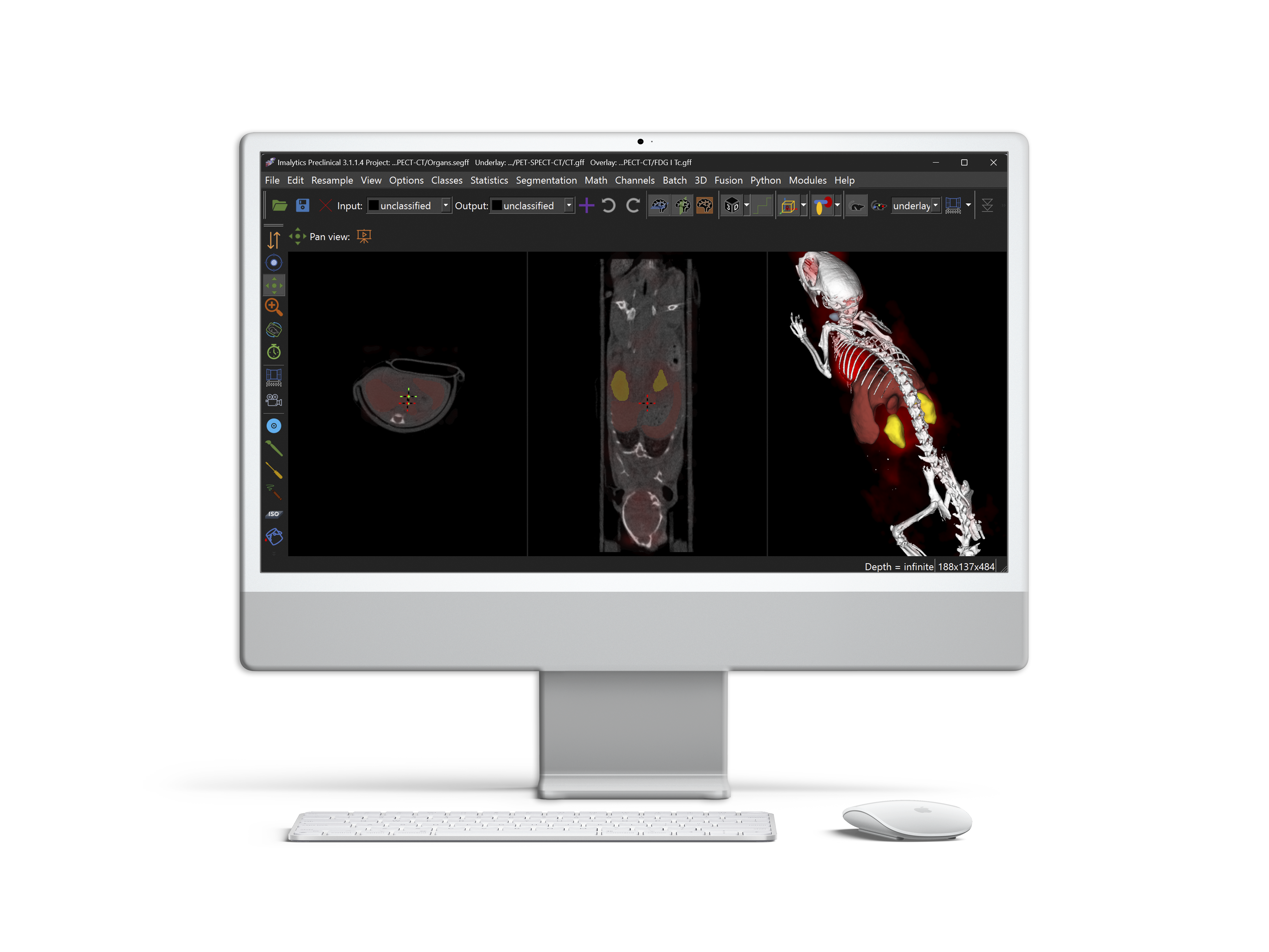
Ready-to-use workflows
  • check-white Increased reproducibility
  • check-white Efficient image analysis
Book a free Demo
banner-1-new

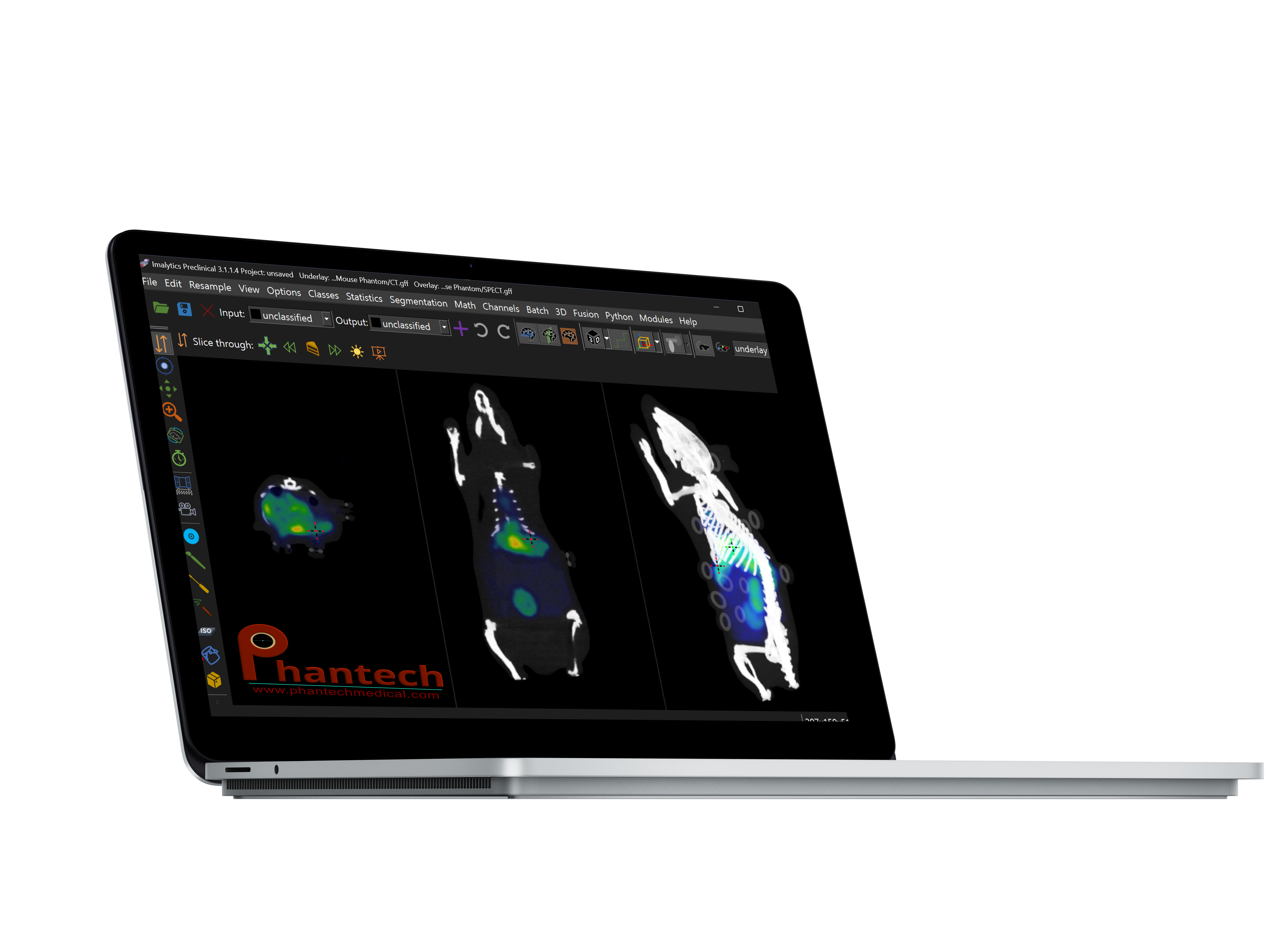
Evaluate System Performance with Automated Phantom Analysis

  • check-white Automated analysis for compatible Phantech phantoms within seconds
  • check-white Assessment for homogeneity, resolution, noise and fusion accuracy
  • check-white Daily Quality control
Book a free Demo
banner-1

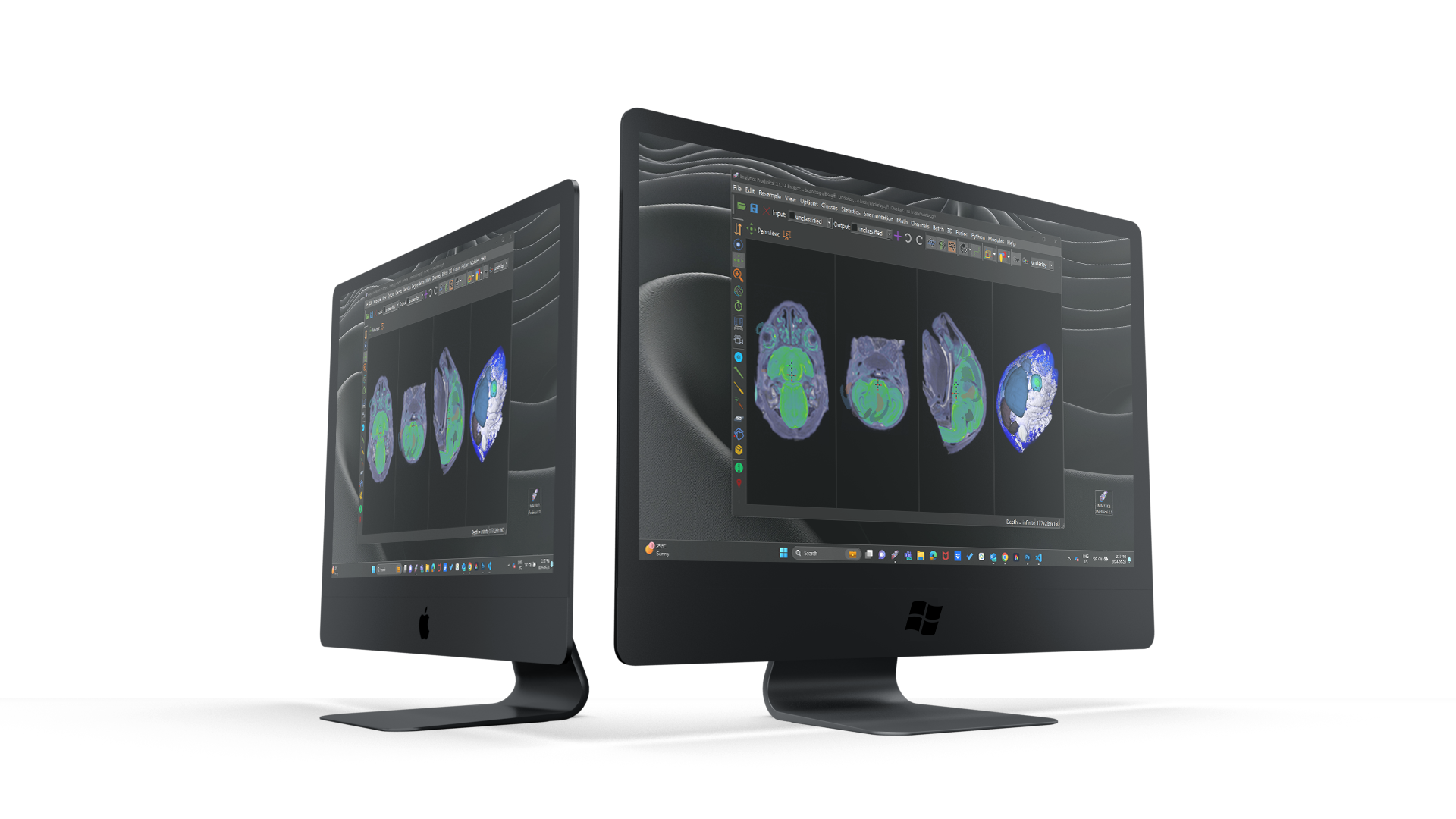
Powerful Platform Independent Software

  • check-whiteLeverage the fastest GPU processing on powerful workstations or benefit from high compatibility for image analysis on any device.
  • check-white Available for Windows, MacOS, Linux, or over the cloud on a powerful remote workstation
  • check-white Affordable and scalable licensing schemes
Book a free Demo
banner-3-new

Explore Advanced Features

Maximize Research Potential with Innovative Features

card-1

Segmentation

Imalytics Preclinical enables fast and easy automatic, semi-automatic, or manual segmentations of phantoms, regions of interest (ROI), or complete organs.

card-1

Multimodal Data

One of our main key expertise lies in the fusion, processing, and analysis of multimodal image datasets such as those issued from a combination of PET, SPECT, CT, MRI, or any other 2D, 3D, 4D or 5D modality.

card-1

Quantification

Quantify signal intensities in phantoms, ROIs, organs, or the whole object using the batch function of Imalytics Preclinical.

card-1

Dosimetry

Deliver precise radiation dose measurements and analysis. Rapidly obtain voxel-based predicted dose measurements, effective dose measurements and dose rate maps for any of our 25+ supported isotopes.

card-1

Bone Analysis

Analyze different bone structures and features by quantifying new bone formation, calcification and porosity while leveraging rapid 3D segmentation tools.

card-1

Python Extension

Perform advanced computations, create custom visualizations, and streamline complex tasks, ensuring your research is both flexible and efficient.

Are you in search of a particular feature? We've
got you covered!

Book a free Demo
logo

Hear From Some of Our Users

Trusted by Leading Experts for Advanced Research Solutions

"The software is easy to learn and provides ready-to-use workflows for different applications."

"Imalytics is an optimal solution as the software offers ease of learning, rapid accessibility, and accommodates numerous users."

"The software allows excellent analysis of multimodal imaging data sets of PET, SPECT, spectral CT and for eg. 19F-MRI combined with proton MRI."

"The tools and applications you've developed on Imalytics are incredibly useful, easy to handle, and effective."

"Imalytics is fast, easy to use and has great support. Great thanks to everyone at Gremse-IT!"

“We have found the Imalytics software to be easy to use and the support from Gremse IT is excellent. We were very impressed with the recently released voxel-based dosimetry module”

Comparison

Imalytics Preclinical in comparison

In-house Developments logo
Thoroughly tested solution cross check
Tutorials & Example Data cross check
Multi-Modal Analysis cross check
Standardized workflows cross check
Available technical support cross check
Continuous updates cross check
Used by 100+ Imaging Cores cross check

Our Process

Get insights about our Process

Live Software Demo

Using your data to understand your specific requirements.

01.

Guided License Selection

Availabe trial license to explore Imalytics.

02.

Support & Onboarding

Product onboarding and constant support.

03.

Request a free demo of Imalytics Preclinical now!

Book a free Demo
logo

Questions and Answers

Frequently Asked Questions

Our image analysis software is not just cost-effective but also user-friendly, offering rapid, automated, and standardized image evaluation that enhances result quality and significantly improves throughput. Allow us to demonstrate its efficacy through a free demo.

Our software is designed to integrate into various workflows, providing centralized data analysis and encouraging collaboration thanks to its uniformity and compatibility with diverse imaging data. Additionally, it can be customized through our Python module, allowing for personal adjustments.

Absolutely. Our platform is intuitively designed, and our comprehensive tutorials minimize the learning curve. Your team can focus on their research, as the software accelerates and simplifies the analysis process.

Our software complements and enhances your existing methods, providing reproducible and standardized data analysis, validated by the community. By integrating your methods via our Python module, you can boost efficiency and broaden the applicability of your innovations.

While free options provide basic features, our paid solution delivers a comprehensive, sophisticated analysis platform. You gain access to advanced analytical tools, our professional support team, regular updates, and a community contributing to the software's continual development.

MILabs

  • U-CT
  • U-SPECT
  • U-PET
  • U-OI

Molecubes

  • CT: X-CUBE
  • PET: ß-CUBE
  • SPECT: y-CUBE

Revvity (formerly PerkinElmer)

  • CT: Quantum GX
  • Optics: IVIS Spectrum, FMT

MRSolutions

  • MRS*SPECT/CT-MR

Bruker

  • CT: Skyscan 1176, 1272, 1276, 1278
  • MRI: 7T

Siemens

  • Inveon CT
  • Inveon PET

Visual Sonics

  • US: Vevo 770
  • Vevo 2100

SOFIE

  • GNEXT PET/CT

Aspect

  • MRI

Philips

  • MRI: 3T Achieva

Mediso

  • nanoScan PET
  • NanoSpect/CT and Tera-Tomo 3D SPECT
  • nanoScan CT
  • nanoScan MRI

Inviscan

  • IRIS PET-CT
  • IRIS CT
  • IRIS XL PET-CT

GE Healthcare

  • CT: Lotus

CT imaging GmbH

  • Tomoscope Duo

TriFoil Imaging

  • Triumph II small animal PET/SPECT/CT

Magnetic Insight

  • Momentum (MPI)
  • Momentum CT (MPI-CT)

NeoScan

  • NEOSCAN N60
  • NEOSCAN N70
  • NEOSCAN N80

Comet Yxlon GmbH

  • Cheetah EVO

SCANCO Medical

  • vivaCT 80
  • XtremeCT II
  • µCT 45
  • µCT 50
  • µCT 90
  • µCT 100
  • µCT 100HE

Xstrahl

  • SARRP
  • SARRP Beamline
  • XenX

United Imaging

  • uBioEXPLORER
  • µMR 9.4 T

Bioemtech

  • β-eye
  • γ-eye
  • φ-eye

Sedecal

  • SuperArgus PET/CT

And many more systems that export image data in standardized file formats, e.g. Nifti, DICOM, or GFF.

The following file formats are supported:


  • Analyze (.hdr/.img)
  • Nifti (.nii/.hdr)
  • MicroPET (.hdr)
  • Vevo770 (.vsi)
  • Vevo2100 B-Mode, NonLinear, Linear (.bimg/.animg/.nbimg)
  • CT-Imaging Files (.mif/.mifx)
  • TIFF (.tif/.tiff)
  • MetaImageHeader (.mhd)
  • Sparse MHD (.sdr)
  • PAR/REC (.PAR)
  • NRRD (.nrrd)
  • NRRD Header Files (.nhdr)
  • PNG (.png)
  • JPEG (.jpg)
  • SkyScan Recons (.log)
  • VFF files (.vff)
  • VOX files (.VOX)
  • Bitmap files (.bmp)
  • Aspect files (.dat)
  • CT Projections (.ori)
  • DICOM (.dcm)
  • Grace File Format (.gff)
  • Super marty View Files (.smv)
  • Interfile (.hdr)